print, etching, engraving
# print
#
pen illustration
#
pen sketch
#
etching
#
figuration
#
ink drawing experimentation
#
genre-painting
#
history-painting
#
academic-art
#
nude
#
engraving
Dimensions: height 140 mm, width 80 mm
Copyright: Rijks Museum: Open Domain
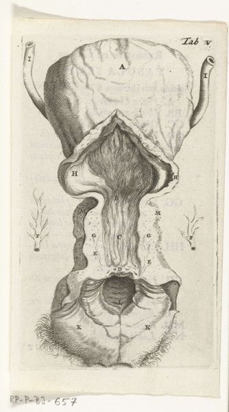
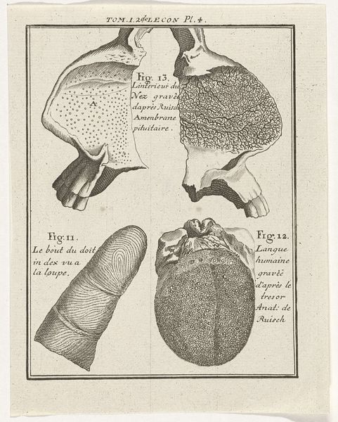
Editor: Here we have Hendrik Bary's "Prints of the Female Reproductive Organs" from 1672. It's an etching or engraving...I'm struck by the clinical detail, yet also the almost baroque way the form is presented. What layers of interpretation do you see at play here? Curator: The image presents us with more than just clinical information. It embodies centuries of cultural memory surrounding the female body. Consider how knowledge was disseminated in this period; this print is not just anatomical, it is performative. Editor: Performative? Curator: Yes. Visual representations shape understanding. How does the careful, almost reverential depiction of these organs influence perceptions of womanhood, reproduction, and perhaps even morality during this era? Are we meant to see clinical detachment or perhaps something more? Editor: I hadn’t considered that. It’s not simply documenting anatomy; it’s constructing an idea, imbuing it with value. Curator: Precisely! Etchings like this would have been circulated, discussed, and interpreted in different contexts. Symbols carry echoes of their past. Each line and carefully rendered detail served not only scientific exploration but also cultural pronouncements. The scientific blends with the social. Editor: It’s a fascinating tension, this blend of science and culture. I'm looking at it in a completely different way now. Curator: Visual language and representation offer unique paths into the complex psychology behind past scientific progress, continually evolving. Editor: Exactly. This was an enlightening exploration of the intersection of art, science, and social values. Thanks!
Comments
rijksmuseum over 2 years ago
⋮
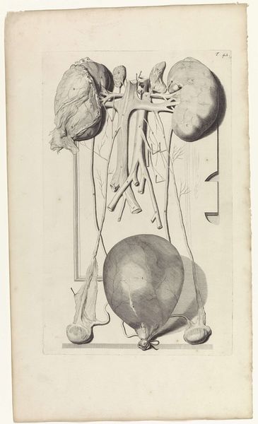
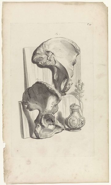
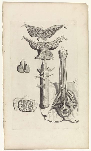
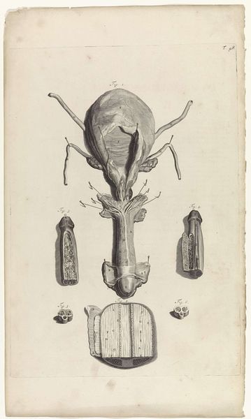
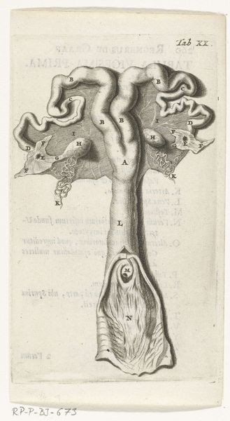
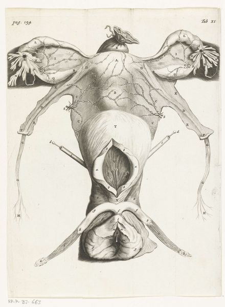
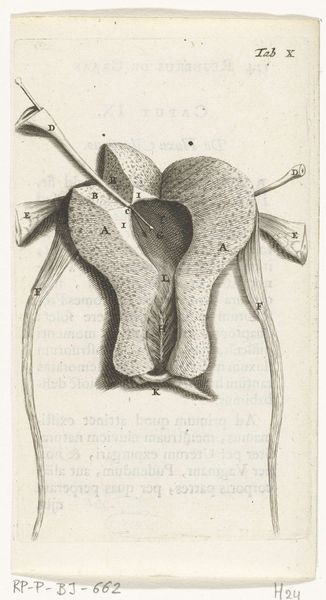
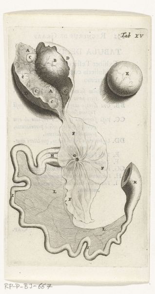
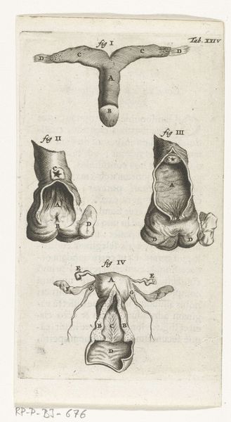
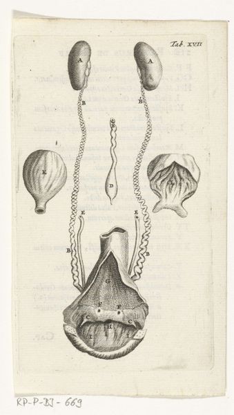
In 1672, physician and anatomist Reinier de Graaf published his De mulierum organis about the female reproductive organs. The book contains detailed prints by Hendrik Bary, among them several of the vagina. De Graaf was the first to conclude that a foetus was the product not just of a man’s seed, but also of a woman’s egg. He discovered what he called blisters, which later became known as Graafian follicles.
Join the conversation
Join millions of artists and users on Artera today and experience the ultimate creative platform.
More like this