print, engraving
# print
#
pen sketch
#
history-painting
#
nude
#
engraving
#
erotic-art
Dimensions: height 140 mm, width 80 mm
Copyright: Rijks Museum: Open Domain
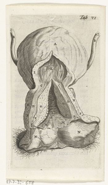
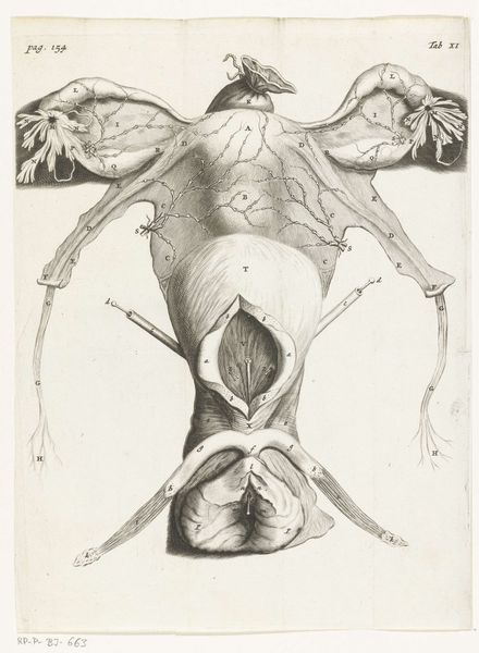
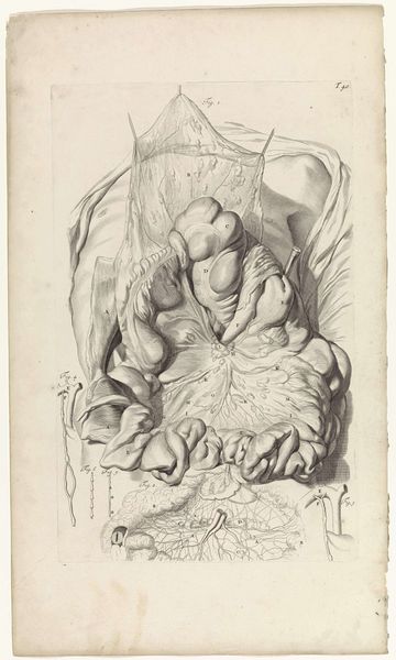
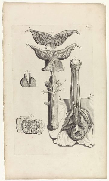
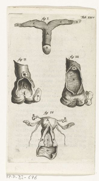
This print of the female reproductive organs was made by Hendrik Bary in the late 17th century, a period deeply engaged with scientific exploration. At first glance, it is a detailed anatomical study, yet it resonates with earlier symbolic representations of the maternal source, as seen in ancient fertility figures. Notice the prominence given to the uterus and ovaries, depicted with careful attention. The depiction of the womb echoes the vessel or container motif found across cultures, symbolizing not just a physical space but a wellspring of life. This connects to earlier images of goddesses and nurturing figures. Consider the cyclical nature of life and death, echoing the constant return of the life force. This anatomical rendering becomes more than clinical; it becomes a vessel of cultural memory, imbued with the potent symbolism of creation and the feminine divine.
Comments
rijksmuseum over 2 years ago
⋮
In 1672, physician and anatomist Reinier de Graaf published his De mulierum organis about the female reproductive organs. The book contains detailed prints by Hendrik Bary, among them several of the vagina. De Graaf was the first to conclude that a foetus was the product not just of a man’s seed, but also of a woman’s egg. He discovered what he called blisters, which later became known as Graafian follicles.
Join the conversation
Join millions of artists and users on Artera today and experience the ultimate creative platform.
More like this