#
toned paper
#
light pencil work
#
quirky sketch
#
pencil sketch
#
personal sketchbook
#
ink drawing experimentation
#
sketchbook drawing
#
watercolour illustration
#
sketchbook art
#
watercolor
Dimensions: height 140 mm, width 80 mm
Copyright: Rijks Museum: Open Domain
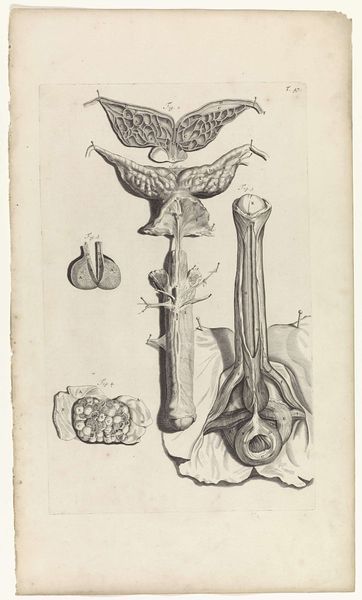
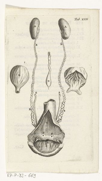
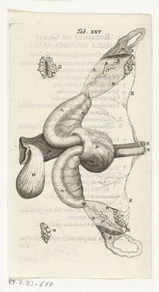
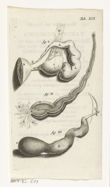
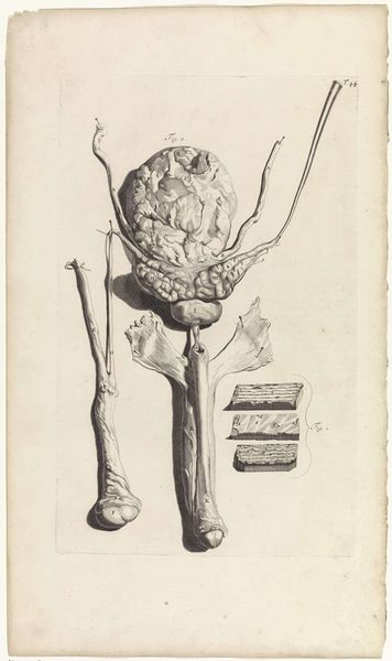
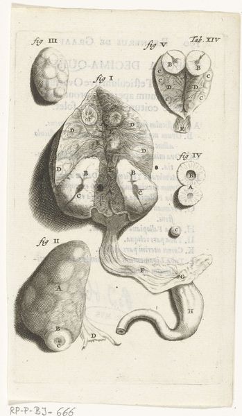
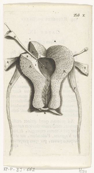
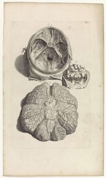
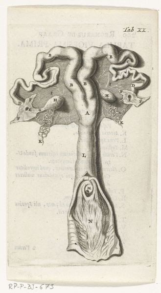
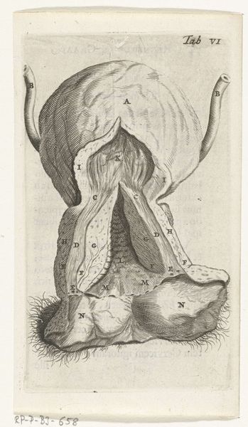
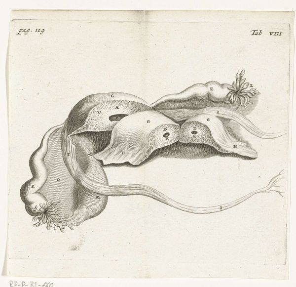
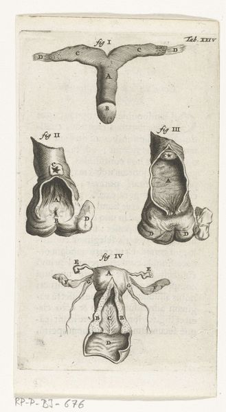
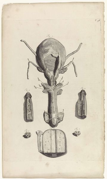
This print of the female reproductive organs was made by Hendrik Bary in the late 17th century. During this time, scientific understanding was intertwined with social beliefs about gender, often reinforcing patriarchal views. This image, with its detailed depiction of the female anatomy, would have been used for medical study, yet also reflects a broader cultural gaze on women's bodies. The clinical representation here is not neutral; it's made by a male artist in a society where women's bodies were often seen as objects of mystery and control. The print invites us to reflect on how medical and scientific illustrations can perpetuate gendered power dynamics, shaping not only medical knowledge but also societal attitudes. Consider how this image might have influenced perceptions of women's health and identity during that era. It reminds us to think critically about the historical contexts that influence how we understand the body.
Comments
rijksmuseum over 2 years ago
⋮
In 1672, physician and anatomist Reinier de Graaf published his De mulierum organis about the female reproductive organs. The book contains detailed prints by Hendrik Bary, among them several of the vagina. De Graaf was the first to conclude that a foetus was the product not just of a man’s seed, but also of a woman’s egg. He discovered what he called blisters, which later became known as Graafian follicles.
Join the conversation
Join millions of artists and users on Artera today and experience the ultimate creative platform.
More like this