drawing, print, etching, ink
#
drawing
#
baroque
# print
#
pen sketch
#
etching
#
pencil sketch
#
ink
#
history-painting
#
academic-art
#
nude
Dimensions: height 140 mm, width 80 mm
Copyright: Rijks Museum: Open Domain
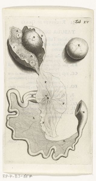
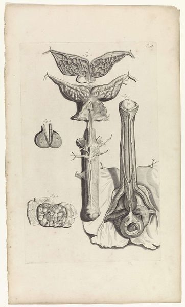
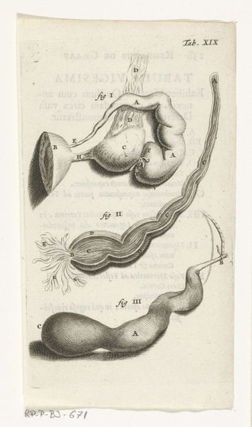
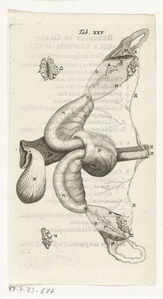
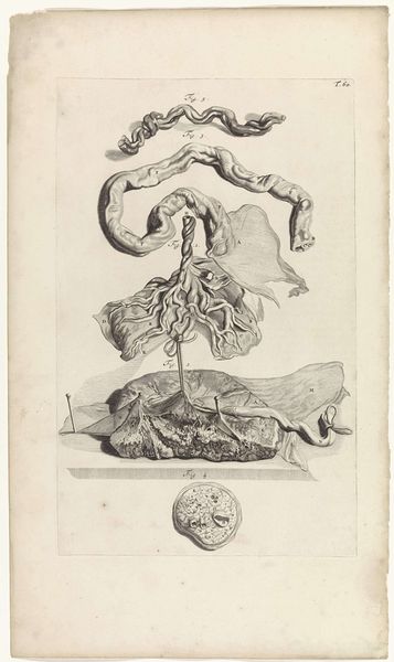
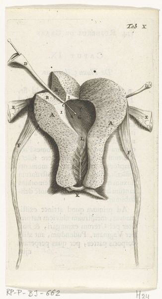
Curator: Here we have Hendrik Bary’s “Prints of the Female Reproductive Organs” from 1672, housed at the Rijksmuseum. It's a fascinating confluence of printmaking, etching, ink and drawing to study an important female bodily structure. Editor: Well, initially, I am struck by the delicate linearity of the engraving, this web of meticulously etched marks coming together into a form that looks so intensely real, while remaining entirely in monochrome. A series of numbered anatomical sketches meticulously layed on a two dimensional plane. Curator: Precisely. Remember that in 1672, depictions of this kind were walking a fine line between scientific illustration and social taboo. It would be viewed either as radical progress or deep sacrilege depending on one's audience. The very act of portraying the female body, normally hidden, was a significant symbolic move. Editor: Yes, there is something fundamentally confrontational about bringing such inner workings into view. What of the individual notations sprinkled across the work, are they meant to be decoded and interpreted? Curator: Most certainly, in addition to labeling elements of female anatomy with the alphabets, this functions as a symbolic map of medical and perhaps philosophical understandings of women at the time. Every marking speaks to a belief system, often reflecting prevailing biases and limitations in knowledge. Editor: And yet, even divorced from their intended meaning, the individual etchings exhibit such clarity in conveying spatial relations with respect to each other; the shadows lend so much dimensionality to them despite the lack of color! Curator: Exactly, this work highlights the evolving cultural relationship between the female body and representation in art, one which blends objectivity with profound social narratives. A very poignant and layered work when thought about it in a wider cultural scope. Editor: I agree. Bary's prints remind us that visual languages can simultaneously clarify and obscure. An analytical lens focused in monochrome.
Comments
rijksmuseum over 2 years ago
⋮
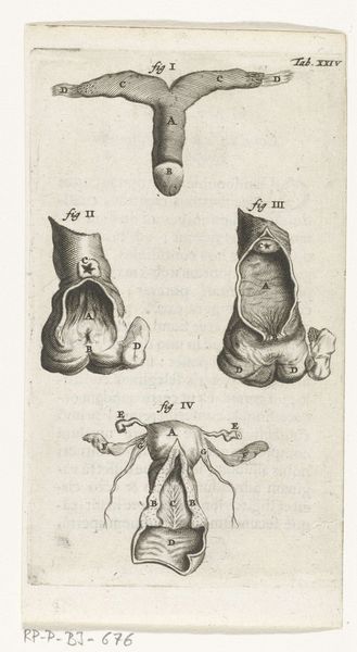
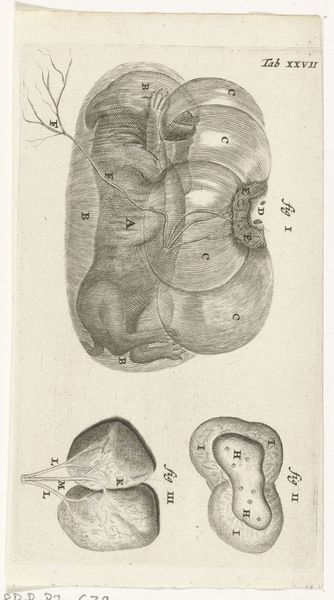
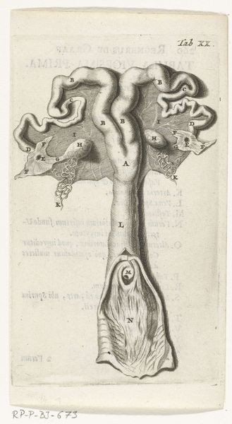
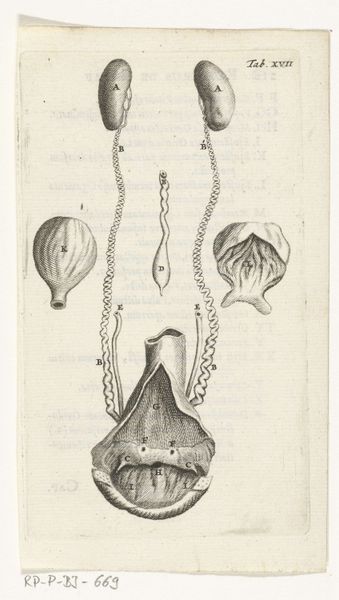
In 1672, physician and anatomist Reinier de Graaf published his De mulierum organis about the female reproductive organs. The book contains detailed prints by Hendrik Bary, among them several of the vagina. De Graaf was the first to conclude that a foetus was the product not just of a man’s seed, but also of a woman’s egg. He discovered what he called blisters, which later became known as Graafian follicles.
Join the conversation
Join millions of artists and users on Artera today and experience the ultimate creative platform.
More like this