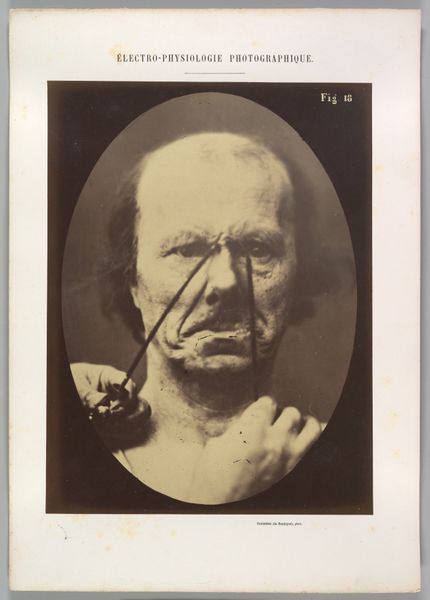
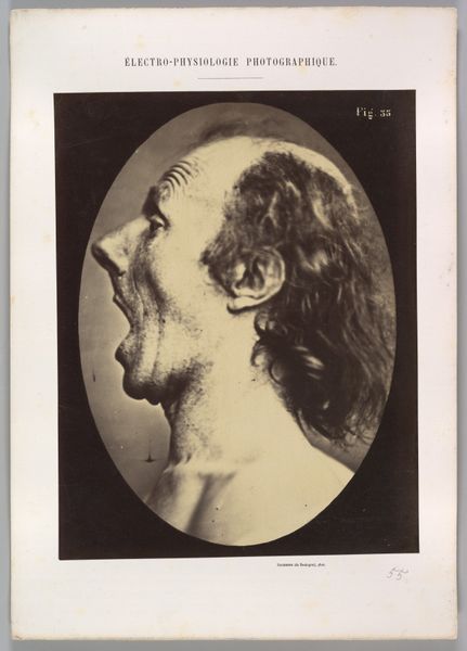
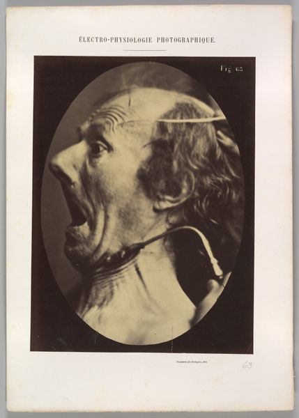
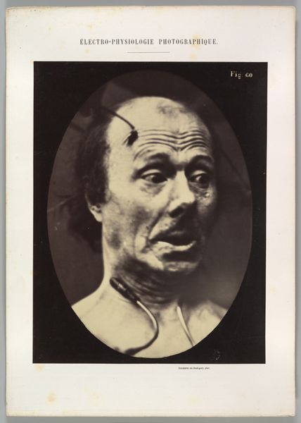
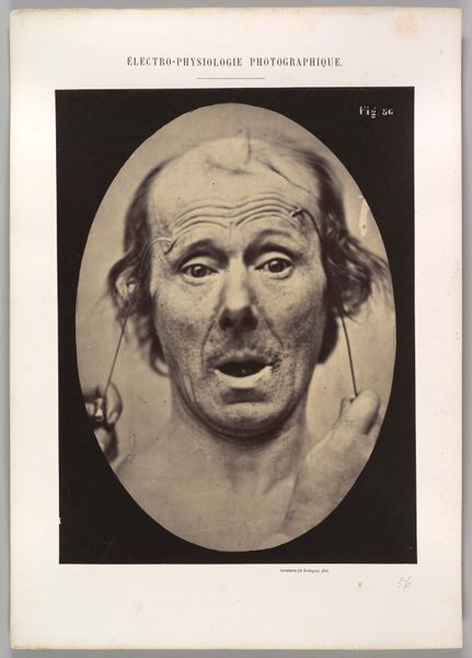
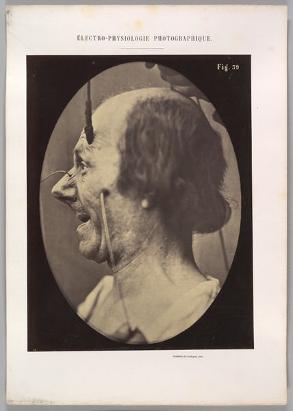
Figure 8: Contraction of the right m. frontalis. 1854 - 1856
0:00
0:00
Dimensions: Image (Oval): 28.4 × 20.2 cm (11 3/16 × 7 15/16 in.) Sheet: 29.8 × 22.9 cm (11 3/4 × 9 in.) Mount: 40.1 × 28.6 cm (15 13/16 × 11 1/4 in.)
Copyright: Public Domain
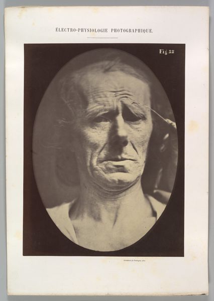
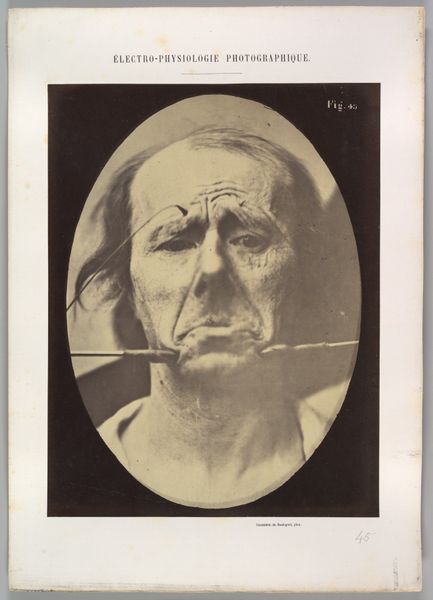
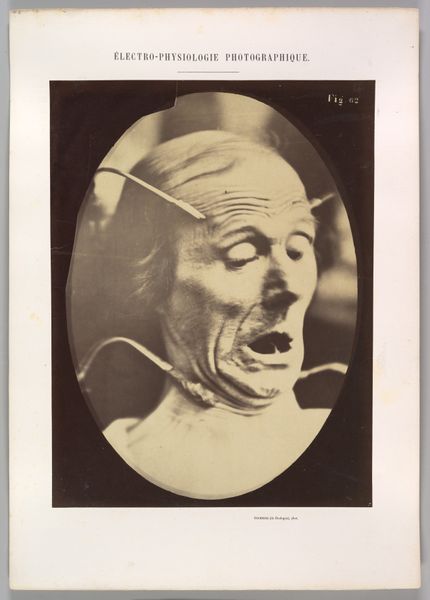
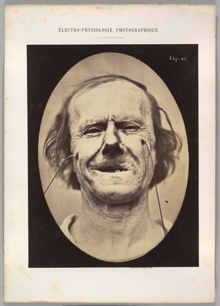
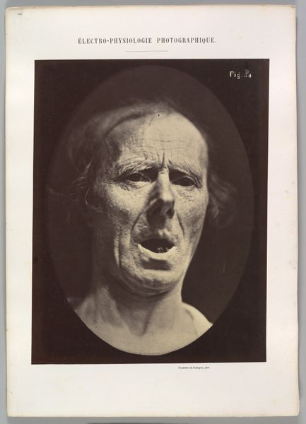
This is a photograph by Guillaume Duchenne, made in France, part of his research into the physiology of facial expression. Duchenne was working at a time when the authority of science was growing and when photography was becoming an ever more important tool for scientific investigation. He used the new medium to record the effects of electrical stimulation on the facial muscles of his subjects. Here, we see an older man with wires attached to his face. The text above the image labels it as showing a contraction of the right frontalis muscle. Duchenne believed that facial expressions were universal and could be mapped scientifically. His work was influential to fields like psychology and even art, informing how artists thought about the representation of emotion. Today, historians of science are interested in Duchenne’s photographs as a source of information about the history of medicine and scientific attitudes toward the body. By looking at these images and the texts that accompanied them, we can learn a lot about the social and institutional contexts that shaped their creation.
Comments
No comments
Be the first to comment and join the conversation on the ultimate creative platform.
More like this