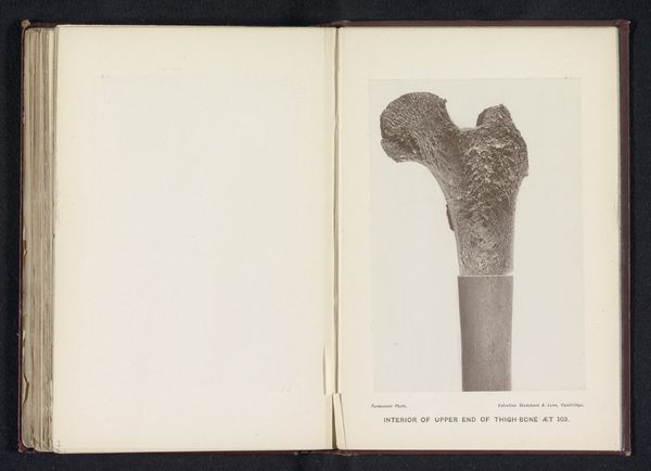
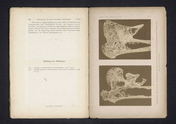
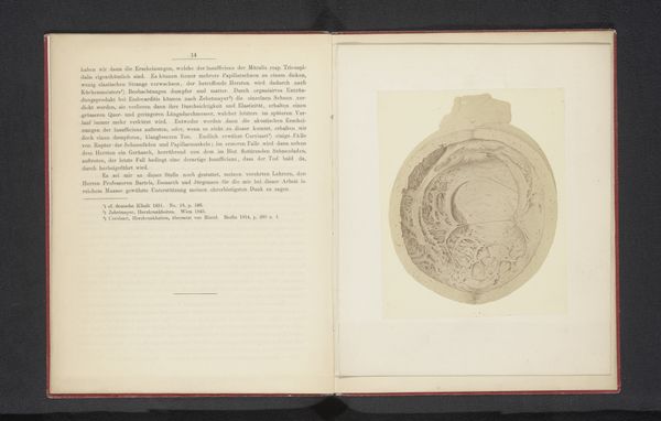
Binnenkant van het bovenste gedeelte van het dijbeen van een volwassene before 1889
print, photography, gelatin-silver-print
portrait
photography
gelatin-silver-print
academic-art
realism
Dimensions: height 142 mm, width 95 mm
Copyright: Rijks Museum: Open Domain
This image shows the interior of the upper end of an adult thigh bone, reproduced as a permanent photo in an anatomical study. Images like these reflect a 19th-century obsession with cataloging and understanding the human body through scientific means. The study of anatomy was undergoing rapid professionalization, and the production of detailed, accurate visual records was central to the institutionalization of this new medical knowledge. How are these images used? Are they for training surgeons or educating the public? Perhaps it served both purposes, shaping public understanding of the body while standardizing medical practices. This wasn't just about science. It was about power – the power to define what is normal, what is healthy, and what is not. Historians of medicine explore archival records, such as medical textbooks and institutional documents, to uncover the social and political contexts in which anatomical knowledge was produced and used. Examining the distribution of these books, how they are presented to the public, and how they might have influenced social behaviors helps us understand their far-reaching implications.
Comments
No comments
Be the first to comment and join the conversation on the ultimate creative platform.
More like this