print, photography, gelatin-silver-print
# print
#
photography
#
gelatin-silver-print
#
history-painting
#
academic-art
Dimensions: height 48 mm, width 75 mm
Copyright: Rijks Museum: Open Domain
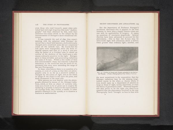
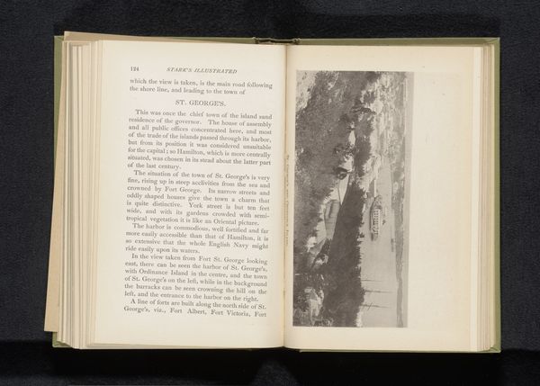
This is a photograph, published at an unknown date by F.R.S. Hicks, showing a portion of a hand with a needle embedded in the thumb. The image is an early example of an X-ray, a technology with transformative implications for medicine and society. The image presents a stark view of the human body, revealing hidden structures beneath the skin. This was revolutionary at a time when medical imaging was in its infancy. Hicks’s photograph reflects a growing interest in scientific advancements and their potential to improve healthcare. Made possible by Wilhelm Röntgen’s discovery of X-rays in 1895, such images quickly moved from physics labs to medical use. They also opened up new ways of seeing, influencing artistic movements like Futurism that sought to capture the invisible forces shaping modern life. To fully understand this image, we can consult scientific literature, medical history, and cultural studies that examine the impact of X-ray technology on society's perception of the body. This photograph reminds us that art and science are intertwined, each reflecting and shaping our understanding of the world.
Comments
No comments
Be the first to comment and join the conversation on the ultimate creative platform.
More like this