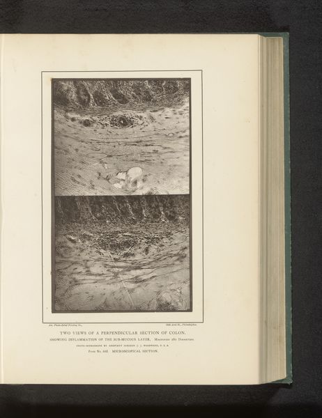
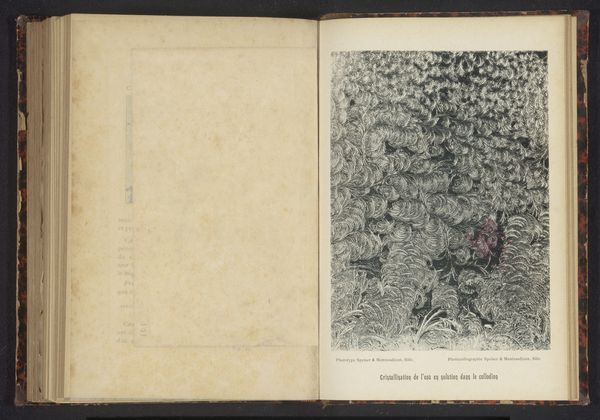
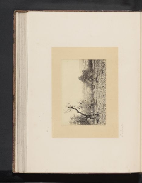
Vergroting van een opgezwollen holte in een stuk dikke darm before 1879
0:00
0:00
drawing, print, paper
#
drawing
#
aged paper
#
homemade paper
#
paper non-digital material
# print
#
sketch book
#
hardpaper
#
personal journal design
#
paper
#
personal sketchbook
#
journal
#
sketchbook drawing
#
history-painting
#
academic-art
#
sketchbook art
Dimensions: height 153 mm, width 117 mm
Copyright: Rijks Museum: Open Domain
Joseph Janvier Woodward, an American army surgeon, made this photo-micrograph of a section of the colon in the 19th century. This was the beginning of modern pathology, where the mysteries of the body were revealed through the microscope and photography. Here, the very structures of human tissue were made newly visible, and disease could be understood in terms of precise physiological changes. This image speaks to an emerging faith in scientific investigation. The image also represents the growth of the American military institution. Woodward worked at the Army Medical Museum, where he applied his knowledge of photography to document specimens collected from Civil War battlefields. These specimens were evidence of the physical and emotional toll of war. By exploring the social context in which this image was made, we can better understand its cultural meaning. We can research archives, institutional histories, and period publications. We can see the image as more than just an illustration of disease. We can view it as a document of scientific progress and institutional growth.
Comments
No comments
Be the first to comment and join the conversation on the ultimate creative platform.
More like this